Global Online Journal of Academic Research (GOJAR), Vol. 3, No. 2, April 2024. https://klamidas.com/gojar-v3n2-2024-05/ |
|||||||||||||||||||||||||||||
|
A STUDY ON THE CADAVERIC ANATOMICAL DIMENSIONS OF THE PTERION IN A NIGERIAN POPULATION: An Important Landmark Guide in Lateral Approach Surgeries*
By Ozor I.I, Nkwerem S.P.U, Iroegbu-Emeruem L, Nnachi S.I, Asimadu I.N, Aroh V.E & Elisa F.I.
Abstract Background: The pterion is a craniometric point near the sphenoid fontanelle of the skull. In neurosurgery, pterional approach is one of the most widely used operative interventions and treatments. The aim of this study is to determine the cadaveric anatomical features of the pterion in a Nigerian population and compare with those of different populations. Materials and method: This is an observational study carried out in the Human Anatomy Departments of Enugu State University College of Medicine, University of Nigeria College of Medicine, Nnamdi Azikiwe University College of Health Sciences, Ebonyi State University College of Medicine and Chukwuemeka Odumegwu Ojukwu University College of Medicine, Nigeria, using fifty cadaveric skulls of both sexes. Oral approval was gotten from the various departments on presentation of letter of introduction from the Department of Anatomy, Enugu State University of Science and Technology, Enugu. Results: The sphenoparietal type of pterion was found to be the most common type (78%) on the right and (74%) on the left, followed by frontotemporal which is (14%) on the right and (18%) on the left; the least common types of pterion is stellate and epipteric with (4%) on the right and the left. Conclusion: Despite the significant variation between the left and right sides of the pterion, the pterions found in cadaveric skulls of Nigerian origin are similar to those of other population.
Introduction The pterion is a reference area in locating the Broca’s motor speech area, anterior pole of the insula, and middle cerebral artery (Sindel et al, 2016). It is also very useful in neurosurgical approaches, especially for anterior circulation aneurysm (Bhargavi, C., Saralaya, V., & Kishan, K., 2011). Furthermore, the pterion which is the weakest areaof the skull, overlies the middle meningeal vessels (Ilknur, Mustafa, & Sinan, 2009), which are the major cause of extradural hematoma in head injuries. (Lama, Mottolese, et al, 2000). Previous cadaveric study around the pterion in SouthEast Nigeria was a single centre study (Ukoha et al, 2013). Besides, the relationship of pterion with the mastoid process, another important neurosurgical landmark, was not explored in the study. This multicenter study is an attempt to determine the cadaveric anatomical features of the pterion in a Nigerian population, compare previous studies in Nigeria and with those of different populations.
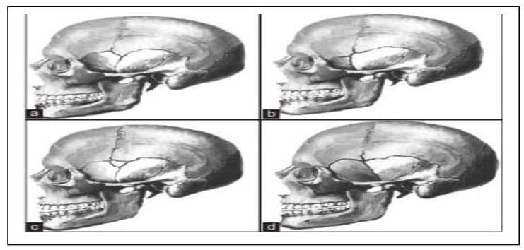
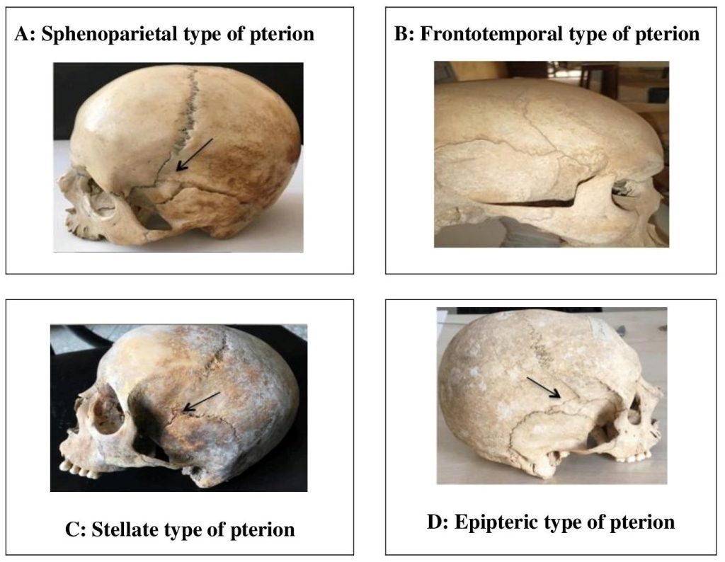
Materials and Methods Study was conducted on fifty human unsexed adult skulls. Dry skulls are taken up for the study as there is paucity of cadavers. Pteria was classified based on Murphy’s classification. Measurements were taken on both sides of the skull from the center of the pterion to the midpoint of zygoma, tip of the mastoid process, Bregma and to the fronto-zygomatic suture using Vernier calipers. Each of the measurements were taken twice then averaged so as to minimize bias errors. Relevant findings regarding various types of pterions and position of pterion were noted. Values were recorded separately on right and left sides and compared. Values were also compared with previous studies for their statistical significance. The Measurement of Pterion This was done in the laboratory, the data collected in this study were statistically analyzed using descriptive statistics like the percentages, mean and standard deviation. To compare between right and left sides, One- Way ANOVA (Analysis of variance) was used. Results In the present study, we found sphenoparietal type of pterion to be the most common type (78%) on the right and (74%) on the left, followed by frontotemporal which is (14%) on the right and (18%) on the left, the least common types of pterion are stellate and epipteric with (4%) on the right and the left. Table 1: The percentage distribution of various types of pterions is shown in the table below.
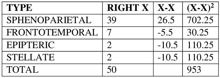
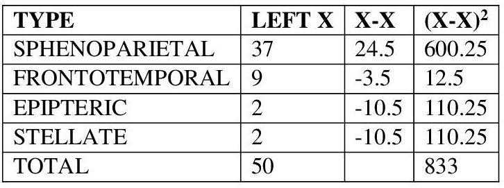
The table below is a comparison of left and right sides using standard deviation Table 2a (right side)
Standard deviation S for the right =17.8 Table 2b (left side)
Standard deviation S for the left= 16. Table 3: ANOVA TABLE
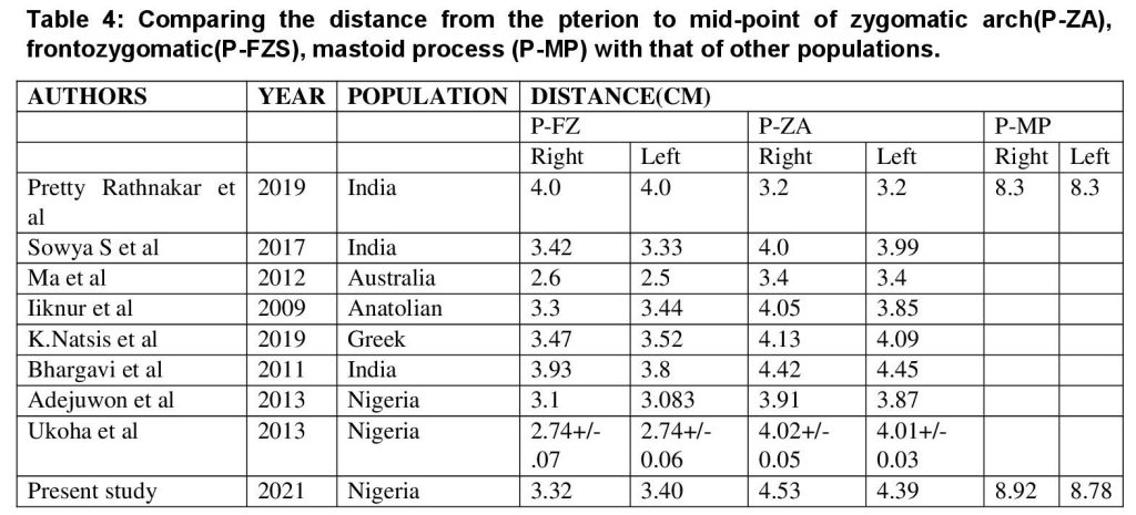
The critical region is F>F (0.5) (3, 4) =F>6.59 Since the calculated f~ ratio (594) is greater than the critical value (6.59) we reject the H0 at α = 0.05 and concluded that the varietal difference is significant. (Note this result is also significant at α = 0.01). Table 4: Comparing the distance from the pterion to mid-point of zygomatic arch(P-ZA), frontozygomatic(P-FZS), mastoid process (P-MP) with that of other populations.
Discussion The predominant pterion found in the present study is the Sphenoparietal type (78%) on the right and (74%) on the left, and this is similar to all other studies summarized in Table 4. In the Asians, the Sphenoparietal pterion frequency ranged between 72% and 93.55%. The Japanese has the highest frequency with (93.5%) followed by the Indians with (72-77%). The Greeks has the lowest frequency (58.4%) followed by the Kenyans (66%). The high frequency of Sphenoparietal pterion could be as a result of evolution (Lui YH, et al 1999), given that it is the commonest type in primates (Ashley- Montagu M. 1933). In the present study, the vertical distance from pterion to zygomatic arch is found to be slightly more on the right side compared to the left side, while the pterion to Frontozygomatic suture was slightly more on the left compared to the right. According to measurement pterion was lying approximately 4.53cm above the arcus zygomaticus on the right side, 4.39cm on the left side and 3.32cm behind the frontozygomatic suture on the right and 3.40cm on the left. A study done in Greek by K.Natsis et al (2019) shows the distance from the pterion to the zygomatic arch to be 4.13cm on the right and 4.09cm on the left, while the distance from the pterion to the frontozygomatic is 3.47cm on the right and 3.52cm on the left. In another study done in India by Bhargavi et al (2011) recorded the distance from the pterion to the zygomatic arch to 4.52cm on the right and 4.45cm on the left, while the distance from the pterion frontozygomatic suture is 3.93cm on the right and 3.8cm on the left. Another study done in Anatolia by Iiknur et al (2009) shows the distance from the pterion to the zygomatic arch to be 4.05cm on the right and 3.85cm on the left, while the distance from the pterion to the frontozygomatic suture is 3.3cm on the right and 3.44cm on the left. Comparing the result from the present study to that of the result gotten from other studies done in different populations it shows that the distance from the pterion to zygomatic arch is found to be slightly more on the right side compared to the left side, while the pterion to frontozygomatic suture is slightly more on the left compared to the right. Conclusion The pterions found in cadaveric skulls of Nigerian origin have the following features when compared to those of other population; Distance from the center of the pterion to mid zygomatic arch and mastoid process was found to be more on the right than the left, from the pterion to the frontozygomatic arch was found to be more on the left than the right and Bregma is equal on both sides. References Adejuwon, S., Olopade, F., & Bolaji, M. (2013). Study of location and morphology of pterion in adult Nigerian skulls. ISRN Anatomy, 1-4. Bhargavi, C., Saralaya, V., & Kishan, K. (2011). Pterion: A site for neurosurgical approach. Int J Biomed Res, 2(12):588-594. Eboh, D., & Obaroefe, M. (2014). Morphometric study of pterion in dry human skull bones of Nigerians. Int J Morphol, 32(1):208-13. Escosa-Bage´, M., Sola, R. G., Liberal-Gonza´lez, R., & Caniego, J. L.-C. (2002). Fusiform aneurysm of the middle cerebral artery. Revista de Neurologia, 655–658. Çimen K., Otag I., & Cimen M. (24th march, 2019). Pterion types and morphometry in middle and south anatolian adult skulls. Rvista Argentina De Anatomica Clinica, 11(1): 8-17. DOI: 10.31051/1852. 8023.v11.n1.21637. Hussain, S., Mavishetter, G., Thomas, S., Prasanna, L., Muralidhar, & Magi, P. (2011). A study of sutural morphology of the pterion and asterion among human Indian skulls. Biomed Res, 22:73 5. Hussain-Saheb, S., Haseena, S., & Prasanna, L. (2010). pterion – Three case reports. Journal of Biomedical science and Research, 2(2): 108–110. Ilknur, A., Mustafa, K. I., & Sinan, B. (2009). A comparative study of variation of the pterion of human skulls from 13th and 20th century anatolia. International Journal of Morphology, 1291–1298. Kaan, C., Ilhan, O., & Mehmet, C. (2019). Pterion types in Anatolian adult skulls. Rev Arg de Anat Clin, 11 (1): 8-17. Lang, J. (1984). The pterion region and its clinically important distance to the optic nerve. 2. Pterion region, distance to the optic nerve, Dimensions and shape of the recess or the temporal pole. Neurochirurgia (Stuttg), 27(2):31-5. Ma, S., Baillie, L., & Stringer, M. (2012). Reappreaising the Surface Anatomy of the Pterion and Its Relationship to the Middle Meningeal Artery. Clin Anat., 25:330-339. Matsumura, G., Kida, K., & Ichikawa, R. (1991). Pterion and epipteric bones in Japanese adults and fetuses, with special reference to their formation and variations. KaibogakuZasshi, 66(5):462-71. Mennan, E., Cem, K., Mehmet, T., Ufuk, C., & Ahmet, H. (2010). Localization of pterion in neonatal cadavers: a morphological study;Surg.RadiolAnat, 32: 545-550. Murphy, T. (1956). The pterion in the Australian aboriginal. Am J Phys Anthropol, 14:225 44. Mwachaka, P., Hassanali, J., & Odula, P. (2008). Anatomic position of the pterion among Kenyans for lateral skull approaches. Int.J.Morphol , 26(4): 931-933. Oguz, O., Sanli, S.G., Bozkir, M.G., & Soames, R.W. (2004). The pterion in Turkish male skulls. Surg RadiolAnat, 26:220 4. http://DOI:10.1007/s00276-003-0210-2 Satheesha, N., & Sowmya, K. (2008). Unusual sutural bones at pterion. International Journal of Anatomical variations, 1: 19-20. Sindel A, Ögüt E, Aytaç G, Oguz N, Sindel M. (2016) Morphometric study of pterion. Int J Anat Res. 4(1):1954-57. Sowmya, S., Meenakshi, B., & Ranganath, P. (2017). Study of pterion: Its clinical and morphological aspects. Indian Journal of Clinical Anatomy and Physiology., 4(2):247-9. Sucharitha, & Roshni, B. (2016). Study of anatomic position of Pterion in dry human skulls in Karnataka. Scholars Journal of Applied Medical Sciences, 4(9B):3272-3276. Ukoha U, Oranusi CK, Okafor1 JI, Udemezue OO, Anyabolu AE, Nwamarachi1 TC. (2013) Anatomic study of the pterion in Nigerian dry human skulls. Niger J Clin Pract.16(3):325–328. [PubMed] [Google Scholar] Vigo V, Cornejo K, Nunez L, Abla A, Rubio RR. (2020) Immersive surgical anatomy of the craniometric points. Cureus. 12(6). Wandee, A., Supin, C., Vipavadee, C., Paphaphat, T., &Noppadol, P. (2011). Anatomical consideration of pterion and its related references in Thai dry skulls for pterional surgical approach. J Med Assoc Thai, (2); 205-214. Williams, P., Bannister, L., Berry, M., Collins, P., Dyson, M., & Dussek, J. (2006). Gray’s Anatomy. 40th edition. In C. Livingstone. Yasargil, M., & Fox, J. (1975). The microsurgical approach to intracranial aneurysms. Surg Neurol, 7 – 14. Yuvaraj, M., & Sankaran, P. (2017). A morphometric study on different shapes of pterion and its clinical significance. Int J Pharm Bio Sc, 8(2):999-1003. Zalawadia, A., Vadgama, J., Ruparelia, S., Patel, S., Rathod, S., & Patel, S. (2010). Morphometric Study of pterion in Dry Skull Gujarat Region. Njirm, 1:25 9. * This is a revised version of an article of the same title published by the authors in 2023.
|
|||||||||||||||||||||||||||||
